Vessel Analysis Assistant
Upload an image and optionally tell the assistant what you would like analyzed.
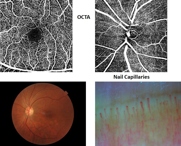
Upload a fundus, OCTA, nailfold, SLO, or other vessel-containing image to begin. I support five image-analysis workflows: (1) fundus photographs—abnormalities,retinal landmarks and vessel measurements; (2) macular OCTA—FAZ and vessel- density measurements; (3) optic-disc OCTA—peripapillary vessel measurements; (4) nailfold capillaroscopy—capillary segmentation and measurements; and (5) granular RGC images—cell segmentation, counting, and size measurements.
Example vessel analysis
This example shows how the uploaded image and vessel segmentation result will be presented after analysis.
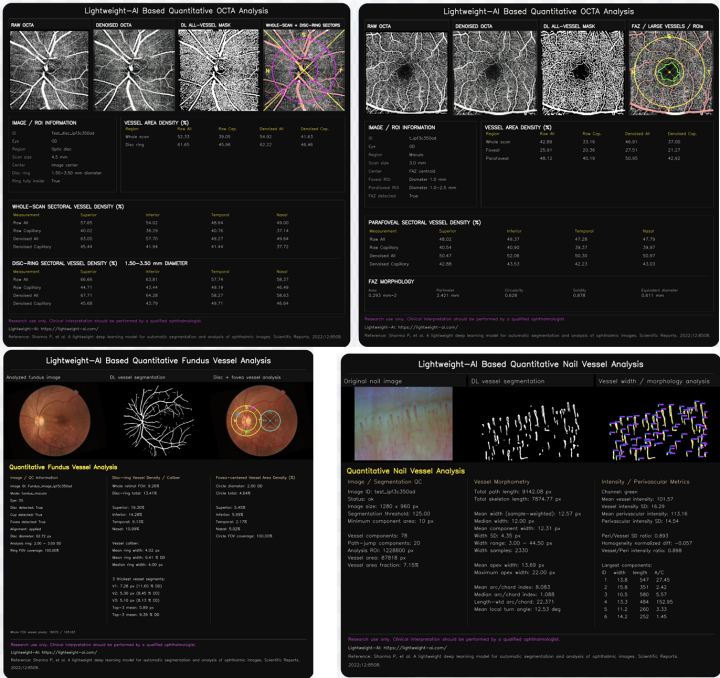
Example analysis
Analyzing image
Processing information is shown temporarily. The final result and report will replace this view when analysis finishes.